Diagnostic Imaging Guide
Learn what diagnostic imaging is and how X-rays, CT scans, MRIs, and ultrasounds help doctors diagnose and treat medical conditions accurately and efficiently.
Medical care today relies heavily on technology that allows doctors to see inside the human body without surgery. This is where diagnostic imaging plays a critical role. From identifying injuries to detecting diseases early, imaging tests help healthcare providers make accurate and timely decisions about patient care.
Understanding the different types of diagnostic imaging can help patients feel more informed and comfortable when their doctor recommends a scan or imaging procedure.
Diagnostic imaging refers to a range of medical tests that create pictures of the inside of the body. These images help doctors diagnose conditions, monitor treatments, and evaluate injuries or abnormalities.
Instead of relying only on external symptoms, imaging allows healthcare providers to look beneath the surface and get a clearer understanding of what is happening internally.
X-rays are one of the most common and widely used imaging tools. They use a small amount of radiation to create images of bones and certain internal structures.
They are often used to detect fractures, infections, and abnormalities in the chest or skeletal system.
CT scans combine X-ray images taken from different angles to create detailed cross-sectional views of the body.
They provide more detailed information than standard X-rays and are commonly used to evaluate internal injuries, organs, blood vessels, and complex conditions.
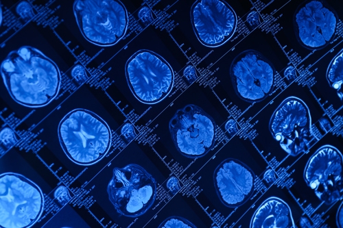
MRIs use powerful magnets and radio waves to produce detailed images of soft tissues in the body.
They are especially useful for examining the brain, spine, joints, and muscles without using radiation.
Ultrasound imaging uses sound waves to create real-time images of organs and structures inside the body.
It is commonly used during pregnancy, as well as to evaluate organs such as the heart, liver, kidneys, and gallbladder.
Diagnostic imaging allows healthcare providers to detect health issues early, often before symptoms become severe. Early detection can lead to more effective treatment options and better patient outcomes.
It also helps doctors monitor ongoing conditions and evaluate how well treatments are working over time.
Most imaging procedures are quick, non-invasive, and painless. Depending on the type of scan, you may be asked to lie still while a machine captures images of the targeted area.
Some tests may require preparation, such as avoiding food or drink beforehand or wearing loose clothing without metal objects.
Your healthcare provider will explain the process before your exam so you know exactly what to expect.
Diagnostic imaging is generally considered very safe. Some procedures, like X-rays and CT scans, use small amounts of radiation, but these levels are carefully controlled to minimize risk.
Other imaging methods, such as MRI and ultrasound, do not use radiation at all.
Diagnostic imaging has become an essential part of modern medicine. It supports nearly every medical specialty, from emergency care to cardiology and orthopedics.
By providing detailed insight into the body, imaging helps physicians make more accurate diagnoses and develop effective treatment plans tailored to each patient’s needs.
Diagnostic imaging plays a vital role in delivering high-quality healthcare. Whether it is an X-ray for a broken bone, an MRI for joint pain, or an ultrasound for internal evaluation, these tools allow doctors to see what the eye cannot.
With advanced imaging technology available, patients benefit from faster diagnoses, improved accuracy, and more effective treatment options.
![]()
7939 US Highway 165, Columbia, LA 71418
(318) 649-6106